Understanding limited dorsal myeloschisis
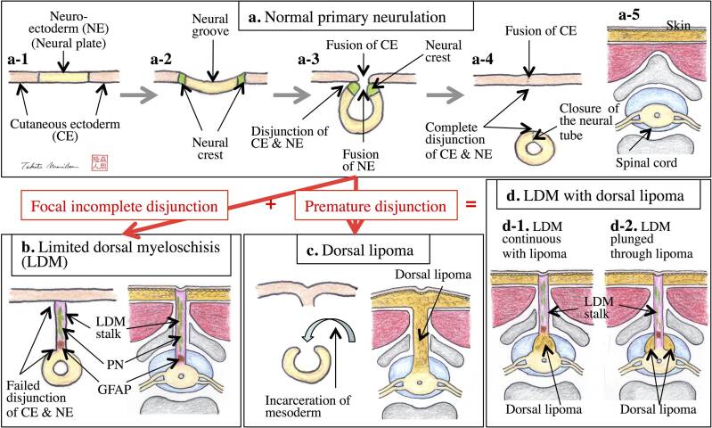
Embryologic origin of LDM
The above figure from Murakami et al. 2020 demonstrates the embryologic origin of LDM. "[LDM is due to] focal incomplete disjunction between the cutaneous and neural ectoderm during primary neurulation, while spinal lipoma of dorsal-type (dorsal lipoma) arises from premature disjunction."
Key clinical features and presentation
Patients with LDM may present with cutaneous findings and neurological deficits
- Cutaneous findings of non-saccular LDM usually include a crater covered with pearly epithelium
- Cutaneous findings of saccular LDM usually include a CSF sac topped by squamous epithelium
- Neurological deficits result from spinal cord tethering and can include weakness, sensory changes, bowel or bladder dysfunction, and gait dysfunction.
Images from Wong, Kan, & Pang, 2020

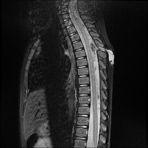
Imaging findings
Per radiopaedia, radiographic features of LDM include the following:
-
a midline skin crater
-
a fibroneural stalk which extends from the skin crater, through a posterior neural element defect (dysraphism), to the spinal cord
-
fibroneural stalk causes tenting/tethering of the spinal cord at the tract-cord union
-
in the saccular subtype of LDM, there is a subcutaneous CSF filled sac coexisting with the fibroneural stalk
Visit the radiopaedia page on LDM to learn more.